
|Articles|October 13, 2020
Cannabis Science and Technology
- October 2020
- Volume 3
- Issue 8
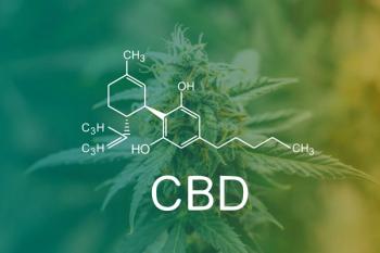
New Study Links Cannabidiol’s Multitudinous Effects to Disruption of Cholesterol Homeostasis
Author(s)Cindy Orser
Op-ed on a new study related to how cannabidiol (CBD) exerts its clinical efficacy.
Advertisement
A significant contribution to our understanding of how cannabidiol (CBD) exerts its clinical efficacy has recently been published online by researchers at the University of Colorado (1). The evidence for how CBD affects more than 20 membrane targets had potentially implicated that the influence of CBD on membrane fluidity was responsible for its pharmacological effects, but the actual mechanism had not been demonstrated until now. The key finding from the multi-omic systems level analysis in human cell lines is that CBD partitions into cellular membranes, which disrupts cholesterol homeostasis along with membrane spanning ion channels and receptors, triggering a myriad of downstream consequences. CBD affected cytosolic calcium, cholesterol transport and storage, cellular energy sensors, and oxidative stress responses in a dose-dependent manner (1). In addition, CBD-exposed cells were sensitive to extracellular cholesterol, which induced cell death, or apoptosis, in a dose dependent manner.
Cholesterol interacts with the fatty acid tails of phospholipids to moderate the biophysical properties of the plasma membrane, reducing fluidity and making the membrane less permeable to small water-soluble molecules that would otherwise freely cross. While cholesterol’s bad press has led to the widespread prescription of statins that block synthesis of cholesterol, it is essential for both membrane permeability and also in metabolism as the starting point for the synthesis of bile salts and steroid hormones including cortisol, estrogen, and testosterone. Cholesterol makes up around 25–30% of the lipid content of the cell membrane and it is the dominant lipid in the brain. Cells either synthesize cholesterol from acetyl coenzyme A (acetyl-CoA) or take it up from the food we eat. While cholesterol deficiency results in several diseases, we are just beginning to investigate and appreciate the regulatory role that cholesterol plays in its relationship to cellular homeostasis, inflammation, and pain much like how we think of CBD today.
Of the proposed list of CBD targets, 22 are membrane-localized channels and receptors. Mutations in one voltage-dependent sodium channel, NaV1.1, are responsible for epilepsy syndromes including Dravet syndrome for which Epidiolex is prescribed (2). Many other proposed targets are calcium channels or receptors, in fact postsynaptic calcium mobilization is the probable mechanism for the anticonvulsant activity of CBD (3). The recent study brings strong evidence to a previously proposed theory that this interaction of CBD with membrane-localized channels is indirect through biophysical alteration of the lipid bilayer with cholesterol as the effector molecule (4,5).
Bioactive Lipids
Growing evidence suggests that pain sensation caused by inflammation is in part regulated by the pro- or anti-nociceptive actions of lipid mediators which include endogenous cannabinoids as well as agonists of peroxisome proliferator-activated receptor-α (PPAR-α), platelet activating factor (PAF), and various products of oxidative polyunsaturated fatty acids (PUFAs) metabolism (6). An increasing number of lipid molecules have been shown to suppress the inflammatory process, restore homeostasis in damaged tissues, and attenuate pain sensitivity by regulating neural pathways that transmit nociceptive signals from the periphery of the body to the central nervous system (7). The endocannabinoids, anandamide and 2-AG, are synthesized locally on-demand to mitigate the effects of exogenous and endogenous pro-analgesic agents by attenuating nociceptor excitability and contrasting local pro-inflammatory signals from another group of bioactive lipids, including membrane cholesterol (7).
Loss of Cholesterol Homeostasis
Let’s take a closer look at how loss of cholesterol homeostasis mitigates a wide range of physiological change. Normally, cholesterol levels are tightly regulated between the plasma membrane (PM) sensors and the endoplasmic reticulum (ER). When cholesterol is depleted, low density lipoprotein (LDL) molecules transporting cholesterol bind to receptors and enter the cell through endocytosis to reach lysosomes where the cholesterol is released from the LDL and delivered to the PM and ER membranes. If there is excess cholesterol, it is removed from the PM and delivered to the ER where it is esterified for storage in lipid rafts and not available for normal cellular processes (8). When this normal feedback inhibition of cholesterol levels is disrupted there is subsequent activation or deactivation of raft-associated proteins, such as death receptor proteins, protein kinases, and calcium channels. Furthermore, increased production of cholesteryl esters, has been reported across human tumors compared with normal tissue.
Cholesterol, Cancer, and CBD
Changes in cholesterol metabolism play a role in carcinogenesis. It is well established that cancer cells have a much higher concentration of cholesterol and the levels of cholesterol determine their resistance to chemotherapeutic drugs (9). This increased capacity to esterify and accumulate cholesterol in tumor cells has been associated with a higher growth rate, suggesting a link between cholesteryl ester production and cell proliferation (10,11). There does not appear to be a correlation with the levels of cholesterol consumed, but rather de novo synthesis from dysregulation of the cholesterol pathway. Several anti-cancer drugs are able to suppress growth and induce apoptosis of tumor cells through alteration of cholesterol containing lipid raft contents via disrupting lipid raft integrity and thereby sensitizing cancer cells to therapeutics and also influencing cancer drug resistance (12). Similar findings have been reported for cancer cells treated with CBD, essentially robbing cancerous cells of their energy source, cholesterol.
In earlier reporting, primary microglial cells treated with CBD resulted in a concentration dependent induction of apoptosis that was not counteracted by CB1 or CB2 receptor antagonists but was attenuated by a lipid raft disruptor. This finding suggests that CBD induces a pro-apoptotic effect in primary microglia through lipid raft coalescence and elevated expression of GM1 ganglioside, involved in neuron repair, and caveolin-1, a structural component of lipid rafts (13). In the case of unregulated cholesterol accumulation in cancer cells, CBD is eliciting the same apoptotic outcome as anti-cancer drugs without the toxic side effects. Likewise, statins have proven therapeutic in some cancers—including gastric, hepatic, prostatic, and esophageal—through their inhibition of cholesterol synthesis (14).
Cellular Calcium, Cancer, and CBD
CBD treatment was associated with high levels of Ca2+ in the cell (1). An influx of Ca2+ ions can damage cells through disruption of neural circuitry through excitotoxicity which can occur in neurodegenerative diseases or brain trauma such as traumatic brain injury (TBI) or stroke even causing cell death. Cellular calcium homeostasis is necessary for stress responses in metabolic organs including the heart, liver, and adipose tissue. Multiple lines of evidence demonstrate that inappropriate consumption of nutrients and chronic metabolic diseases leads to dysregulation of cytosolic and organelle Ca2+ fluxes and disrupted equilibrium in metabolic tissues and immune cells, which in turn alters organelle homeostasis, signaling pathways and autophagy (15). Cellular Ca2+ homeostasis fails to maintain its adaptive integrity in the face of structural and signaling defects introduced by chronic metabolic challenges whether through over consumption of nutrients or through treatment with CBD.
The effects rendered by CBD treatment cannot escape the interrelatedness of all processes in the human body. In a recent study by Olivas-Aguierre and colleagues (2019) the treatment of acute lymphoblastic leukemia of T-cells (T-ALL) with CBD resulted in mitochondrial dysfunction and cell death by altering their calcium homeostasis, which was directly demonstrated as the inhibitor of mitochondrial Ca2+ uptake protected the T-ALL cells from CBD-induced cell death (16). Importantly, and of even further interest open to speculation, is that healthy, resting T-cells were resistant to the effects of CBD and avoided cell death! These data provide provocative evidence for CBD as a candidate for chemotherapy trials.
CBD-Induced Mitochondrial Dysfunction
We now understand that not only does CBD intercalate into the plasma membrane, disrupting ion channels, but it also activates internalization of PM cholesterol which in turn destabilizes the ability of mitochondria to manage its own calcium homeostasis affecting multiple cellular processes including Ca2+ signaling the downstream cell death. CBD also causes the translocation of other membrane proteins leading to decreased cellular energy (ATP) production. At play here is the translocation of a key mitochondrial membrane protein, hexokinase I (HK1). Loss of functioning HK1 results in the decoupling of glycolysis from mitochondrial respiration through diminished transformation of glucose to glucose 6-P (17). Through multi-omic profiling in combination with fluorescence resonance energy transfer (FRET)-based biosensor screening, the Colorado group’s results confirm an earlier report by Alharris and colleagues (2019) that CBD-induces mitochondrial dysfunction (18).
Mitochondrial dysfunction leading to apoptosis or cell death, reported in the Colorado study, has also previously been shown in pancreatic b-cells with associated insulin resistance prior to cell death a hallmark in type 2 diabetes (19). Findings from another relevant study this time involving neurons and platelet-activating factor (PAF), which is also influenced by calcium levels and PM cholesterol is worth citing here (20). PAF binds to its receptor (PAFR) located in the PM and nuclear membrane which mobilizes Ca2+ and activates numerous signaling pathways including inflammation, which can result in cell death. If cells are treated with statins or CBD to reduce the level of cholesterol, then PAFR is displaced, PAF doesn’t bind to its receptor and neurons become resistant to apoptosis. This is a significant finding in light of another study where deletion of PAFR in a mouse knockout experiment abolished the inflammatory response and neuronal apoptosis caused by TBI (21). As a side note, Etizolam, a benzodiazepine analog, is a PAF-receptor antagonist used to treat anxiety.
Far Reaching Implications
The widespread self-medication with CBD, now accessible in a dizzying array of consumables readily available to the public, has preceded any clear understanding of the mechanisms behind CBD’s apparent benefit across a wide range of ailments. The far-reaching cellular disruptions observed in the Colorado study were corroborated by transcriptomics, metabolomics, and proteomics showing the downstream impact from disruption in cholesterol biosynthetic and regulatory pathways including decoupling of glycolysis from mitochondrial respiration, upregulation in oxidative stress response, increase in Ca2+ cytosolic levels, and the increased production and storage of cholesterol sensitizing cells to apoptosis making cholesterol the effector molecule of CBD across many intracellular pathways rather than any specific target.
The Colorado study has shown that not all CBD effects on cells are beneficial. While there had been earlier reports from human clinical trials of the now Food and Drug Administration (FDA)-approved CBD product, Epidiolex, indicating that CBD treatment could lead to risk of liver injury as indicated by increased levels of aminotransferase activity and inhibition of P-450 (CYP) enzymes (22), the molecular mechanisms of that potential toxicity were not fully understood (23).
It should be mentioned that the Colorado study was carried out on two immortalized human cell lines: SK-N-BE(2) neuroblastoma cells and HaCaT human keratinocyte cells with CBD treatment in the micromolar range which would be less than the prescription dosing of Epidiolex, but probably more than self-administered CBD dosing. Their analyses based on transcriptomics showed that the enriched transcription factors shared oxidative stress as a stimulus, resulting in upregulation of oxidative stress response including fatty acid and cholesterol biosynthetic gene transcripts. It was known that CBD upregulates mRNA transcripts in microglial cells involved in fatty acid metabolism and cholesterol biosynthesis (24). This alteration in fatty acid metabolism was also shown in a mouse chronic alcohol feeding study where CBD reduced the infiltration of liver cells with fat, a distinct benefit (25).
If anything, this study has brought attention to the need for basic research on cannabinoids and the biological systems they effect to clarify why endocannabinoids are synthesized locally on an as needed basis in nanomolar amounts and then are quickly degraded. Fundamentally, the endocannabinoid system presents a unique opportunity to look at how a lack of essential nutrients, specifically lipid-based foods, or an overabundance of low-quality nutrient intake impacts synaptic plasticity and resulting outcomes in physical and mental health.
References
- S.E. Guard, et al.
https://www.researchgate.net/publication/341935024_Multi-Omic_Analysis_Reveals_Cannabidiol_Disruption_of_Cholesterol_Homeostasis_in_Human_Cell_Lines (2020). - C. Dravet, Epilepsia 52, Suppl 2:3-9 (2011).
- N.A. Jones, et al., J. of Pharma. and Exptl. Therapeutics 332, 569-577; DOI:
https://doi.org/10.1124/jpet.109.159145 (2017). - M-R. Ghovanloo, et al J. Biol. Chem. 293, 16546-16558 (2018).
- A.R. Watkins, Channels (Austin) 13, 162–167 (2019).
- M. Amsalem, et al., EMBO J 37, e97349 (2019).
- D. Piomelli and O. Sasso, Nat. Neurosci. 17, 164–174 (2014).
- A. Das, ELife 3, e02882. DOI: 10.7554/eLife.02882 (2014).
- Y. Yamamoto, et al., Biochem. Biophys. Res. Commun. 495, 1292–9 (2018).
- E. Ikonen, Nat. Rev. Mol. Cell Biol. 9, 125–138 (2008).
- S. Dessi, et al., Biochem. J. 321, 603–608 (1997).
- L. Gu, et al., FEBS Press 286:
https://doi.org/10.1111/febs.15018 (2019) - H.-Y. Wu, et al., Glia 7, 1182-90 (2012).
- A. Stryjkowska-Gora, et al., Contemp Oncol (Pozn) 19, 167–175 (2015).
- A. Giordano, et al., Journal of lipid research 54, 2423–2436 (2013).
- M. Olivas-Aguirre, et al., Cell Death and Disease 10, 779–97 (2019).
- L. Berger, et al, J. Gen. Physiol. 29, 379–391 (1946).
- E. Alharris, et al, Ocotarget 10, 45–49 (2019).
- Y.-F. Zhao, et al., Endocrine 37, 76–82 (2010).
- H. Mikashima, et al., Jpn. J. Pharmacol. 44, 387–91 (1987).
- X.J. Yin, Sci. Rep. 7, 40614 (2017).
- M.A. Alsherbiny and C.G. Li, Medicines 6(1), 3 (2019).
- E.A. Thiele, et al., Lancet 391, 17-23 (2018).
- N. Rimmerman, et al., Cellular and Molecular Neurobiology 31, 921–930 (2011).
- Y. Wang, et al., Scientific Reports 7, 12064 (2017).
About the Author
Cindy S. Orser, PhD, is Chief of Science at CLIP Labs in San Diego, California. Direct correspondence to:
Articles in this issue
over 5 years ago
The FDA is Coming…Will the Cannabis Industry Be Ready? Part Iover 5 years ago
Cannabis Cultivation: The New Agriculture Tech Playgroundover 5 years ago
Integrated Data in Cannabis ERP: From Greenhouse to Productionover 5 years ago
How to Rein in Cannabis Industry Energy?over 5 years ago
THC Remediation: A Basic UnderstandingNewsletter
Unlock the latest breakthroughs in cannabis science—subscribe now to get expert insights, research, and industry updates delivered to your inbox.
Advertisement
Related Content
Advertisement


Advertisement
Advertisement